|
...continued from page
2
Many believe the body only begins to mechanically
malfunction when some component is damaged. They miss the
small tolerances necessary for the most efficient and effective
working of this complex a machine. They also miss the importance
of instantaneous transmission of mechanical stress by the
meninges and the fact of true interdependence of all motion
in the body and, most importantly they miss the fact that
bones and other structures can and do get moved in directions
from which the body cannot retrieve them.
Other people do approach structural therapy with the presupposition
that the condition presented can be a distant sequel to a
mechanical pathology elsewhere. Those practitioners have the
idea of slight changes in position causing large changes in
function of the body and they have the thought of interdependence
of motion (holism). However, they most often act and treat
on a local effect basis because the theories on which their
treatment is based have no specific cause consistently and
predictably accounting for ALL the phenomena noticed in the
body.
Via objective analysis of biomechanics using standing and
sitting radiographs of the entire spine on one film or using
two 14"x17" sectionals shot at 72" with the
patient completely relaxed, the practitioner will find a consistent
index and pattern of change in mechanics that can be used
to determine the primary biomechanical pathology(ies) for
which the compensations are generated, resulting in the various
patterns of sequelae named as diseases. Knowing that data,
appropriate application of biostructural treatments can be
instituted. (This can now be done without x-ray.)
Nothing in this presentation should be interpreted to mean
that manipulation of osseous structures is the only, treatment
in these disorders. Depending upon the extent and intensity
of the condition presented or permanent damages developed
as sequelae to the mechanical pathology other biostructural
therapies might be included to allow motion of the structures
into their optimum positions or to provide the support needed
for recovery. There is also the need to develop supports for
those not able to fully recover to maintain and improve the
extent of recovery available to all.
As Breig notes in his comment describing the sequence of
improvements of neurological function in Cervicolordodesis
patients (1), the traditional naming of neurological and spinal
cord disorders "according to début, epidemiology,
acute or chronic nature, etc., does not reflect the histodynamic
causation of the symptoms." That method of naming disorders
has misled practitioners into thinking they have different
etiologies. Breig further notes in that discussion, "It
would be useful if the origin of the tension were stated in
the diagnosis, for then the patient is more likely to receive
the appropriate treatment."
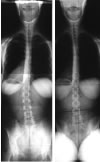
Below are films which demonstrate some of these changes and
a basic explanation.
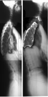
These are the films taken on the same person on the same
day. The person is standing and sitting each picture is one
minute apart. Note some changes on the laterals.
These pictures are not very clear for faster loading of this
page. If you right click on the picture and click open in
another window, you can get a more clear view. Do it for each
picture pair and place them side by side. You can also print
them out and look at the hard copy which will have better
resolution.
Most notable on the lateral view films above
is that the cervical spine is military standing but becomes
normally lordotic sitting. Also, the lumbar spine is in a
hyperlordosis or sway-back standing and military/top half
slightly reversed when sitting. These two curves are supposed
to go in the same direction in a normal person. In this person,
at the time of the x-rays, one is normal in the standing position
or a bit more than normal, while the other is reversed. Sitting
they switch but are still opposite. There is a compensation
mechanism here in which they are working synchronously (changing
instantly in concert with one-another)?
Which one do you treat? How do you treat it, and in which
direction? Does it matter where the patient had pain? Should
you treat at that point? Look below.
These are cutouts of the thoracic spines from the above films.
The sitting thoracic section (middle) has been rotated 12o
counterclockwise so the curves can easily be compared.
How about the thoracic spine? 
In this person the thoracic spine shows very little change
from standing to sitting. It is easy to note that there are
few significant changes from standing to sitting. All the
changes are below the level of the apex of the kyphosis which
is at T10 sitting and above T4 standing. (Apex = furthest
point backward or forward in a curve. Important in analyzing
the point of focus of mechanical stress.)
Also note that the mechanical leverage created by those changes
in the thoracic spine, small as they are, account for the
fact that the lumbar spine becomes hyperlordotic as the cervical
spine loses its lordosis standing while the cervical spine
is not forced to become hyperlordotic in compensation sitting
when the lumbar spine loses its lordosis. Some of the increase
in mechanical stress is taken up by the thoracics.
What about between T5 and the apex of the kyphosis when standing?
The thoracic spine above T10 is not really a kyphosis. It
just drops forward and would probably completely fold forward
if it were not for the ribs. The ribs do not hold the thoracic
spine rigid as so many biomechanical theories state they do.
The ribs provide some support, but not stiffness to the point
of rigidity. The thoracic spine has plenty of motion, especially
in the AP direction.
The thoracic spine is a major compensator of biomechanical
pathology and the most frequently injured portion of the column
in P-to-A traumas such as automobile collisions.
That fact is not yet well documented or researched because
not many doctors seem to notice. The reason might be that
humans, in the standing position, use the large muscles attaching
from the legs to the pelvis and spine to flex the pelvis and
twist the lumbar spine into a hyperlordosis forcing the trunk
backward to balance the collapsed thoracic spine. That makes
the collapse of the thoracic spine less noticeable in the
standing position as a biomechanical event because a pseudokyphosis
is created by the compensation of the lower thoracic spine
(canted posterior) and the portion of the thoracic spine above
the standing apex falling anterior.
Why do the head and neck not fall anterior? They do but not
completely. The effect of the meninges (Breig) and the leverage
effect of the change in curve between the upper thoracic and
cervical spine hold the neck and head up as much as possible
just as the lumbars force the thorax posterior. What is often
noted as normal is not even close to the optimum position.
The variations from normal account for thoracic outlet syndrome
including vascular and neurological signs as well as the many
other symptoms and effects noted in this patient.
Common in the literature of radiology describing the various
radiographic measurements of posture is the comment that such
and such a range is normal. However, there is no specific
correlation between findings outside of the range and symptomatology
in the patient. The reason for this is stated at the outset
of this presentation. The data of these measurements is not
correlated with other mechanical data from the entire spinal
column-pelvis to determine relative changes. With data from
the entire spinal column-pelvis specific correlations between
measurements and would quickly be determined.
The basic correlation is the determination of the lateral
direction of that patient’s primary biomechanical pathology
at that time.
The hypothesis for finding that, simply, follows this line.
When standing, the body can arrange the bones of the lower
extremities and use the contraction of the muscles of the
lower extremities to twist the pelvis and spinal column into
a compensated position.
When one sits with the feet flat on the floor before them,
the use of the lower extremities and muscles are reduced.
They cannot twist and pull enough to compensate as well so
the collapse of the thoracic spine above a given point becomes
more evident. Why say "more evident"? Shouldn’t
the phrase just be evident? No, the collapse is noticeable
standing if one knows what to look for.
After reading this you can probably find the flat spot in
the lower thoracic curve (T12,11,10) above which the thoracic
spine collapses even in the standing view. Go back to that
film and check..
Comparing the standing film to the sitting one can predict
the sites of pain via mechanical stress analysis and correlation
of intensities with direction of mechanical stress. Also,
the vertebrae in need of treatment can be determined since
one can determine which are in flexion and unable to be repositioned
by the body.
On the other hand, once one has x-rays comparing the pelvic
tilts one can determine to which direction the body is falling
due to inadequate bone leverage resulting from the biomechanical
pathologies (people do stay upright because of muscle power
but it is less necessary to use the muscles as the bones become
more optimally positioned for leverage). Using that information,
one can observe the body response to simple physical testing
and determine which vertebrae to treat and which to ignore
on any given day be they at the level of another type of pathology
or not.
Important is that the segments malpositioned but not to be
treated is determined. This is a vital determination because,
though they may also be out of optimal position and may be
at the site of mechanical stress causing other damage, those
segments are out of position to compensate other malpositioned
vertebrae and actually support the body. Changing their position
can change their
continued on page
4 ...
top
|


